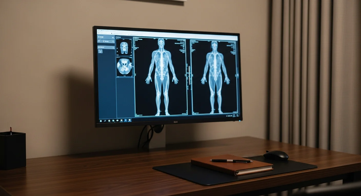
MR angiography is an MRI tuned to show blood vessels, and it maps arteries without radiation and often without any injected contrast. Fishtown Medicine chooses MRA over CT angiography when a person needs repeat imaging over years, has kidney disease that makes iodinated contrast a problem, or is young enough that cumulative radiation matters. CT angiography stays the better choice in emergencies, for heavily calcified arteries, and for the coronary arteries.
TL;DR: An MRA is an MRI tuned to show blood vessels rather than tissue, and its two advantages are that it uses no radiation and often needs no injected contrast at all, since flowing blood produces its own signal. That combination makes it the natural choice when arteries need watching over years rather than looking at once: a small aneurysm being followed, a young person with unexplained high blood pressure, or someone whose kidneys make iodinated contrast a bad idea. CT angiography is faster, more available, and better where calcium is heavy or the question is urgent, so the choice between them is usually about your situation rather than about which machine is better. The sections below are organized by which arteries are in question.
What is an MRA, and how is it different from a regular MRI?
An MRA is the same scanner and the same visit as an MRI, running sequences designed to make blood stand out from everything around it. Where a standard MRI is tuned to distinguish grey matter from white matter or cartilage from bone, an MRA is tuned to render the inside of arteries as a map you can rotate and follow.
There are 2 ways to do it, and which one you get changes whether you need an injection. Time-of-flight MRA uses the movement of blood itself: the scanner saturates the signal from stationary tissue, so blood flowing into the slice arrives unsaturated and appears bright against a suppressed background. Nothing is injected. That is how most brain MRAs are done, and it is why a scan of your intracranial arteries can avoid needles altogether. Contrast-enhanced MRA injects gadolinium and images while it passes through the vessels of interest, which gives cleaner pictures over longer stretches of artery and handles the chest, abdomen, and legs better than time-of-flight can.
Both take roughly 20 to 45 minutes, both are loud, and both carry the same implant and claustrophobia considerations as any MRI, which the MRI guide covers in detail.
Brain and neck arteries
Intracranial MRA is the standard non-invasive way to look at the arteries inside the head, and it is usually done without contrast.
The most common reason to order one in a person without symptoms is a family history of brain aneurysm. Screening is generally considered when someone has 2 or more first-degree relatives who have had an aneurysmal subarachnoid hemorrhage, or when they have autosomal dominant polycystic kidney disease, which carries a higher rate of intracranial aneurysm. A single affected relative usually does not meet the bar, and that conversation is worth having properly rather than deciding by anxiety in either direction.
The other reasons are symptom-driven: a stroke or transient ischemic attack where the question is whether a vessel is narrowed or occluded, an unusual headache pattern that raises concern about a dissection, or a pulsatile noise in one ear. A sudden, severe headache that peaks within seconds is a different situation and belongs in an emergency department, since that presentation is evaluated with CT first and does not wait for an outpatient MRA.
For the neck arteries, MRA competes directly with carotid ultrasound, and the division is practical. Ultrasound is cheaper, faster, and perfectly adequate to grade narrowing in the accessible part of the carotid, so it goes first. MRA earns its place when the ultrasound is technically limited, when the question involves the vertebral arteries or the segments running up inside the skull base that ultrasound cannot reach, or when a dissection is suspected, since MRA with specific fat-suppressed sequences can show blood within the vessel wall itself.
Renal arteries
Renal MRA images the arteries feeding the kidneys, and the clinical question is nearly always high blood pressure that does not behave the way ordinary hypertension does.
Two patterns raise it. The first is a young person, often a woman under 50, with hypertension that appeared without the usual metabolic company, where fibromuscular dysplasia produces a characteristic beaded appearance in the renal artery. The second is resistant hypertension in an older person with atherosclerosis elsewhere, where the question is whether a narrowed renal artery is driving the pressure. Other clues include a sudden worsening of previously controlled blood pressure, a rise in creatinine after starting an ACE inhibitor or ARB, or an unexplained size difference between the 2 kidneys. Most of the time a blood pressure medication that stops working has a more ordinary explanation than a narrowed artery, and working through those first is what keeps this from becoming an imaging problem.
Before ordering imaging, it is worth confirming that the workup for secondary hypertension has been done in the right order, because renal artery narrowing is not the most common curable cause. Primary aldosteronism is more common and is missed far more often, and the primary aldosteronism guide covers why that screen should usually come first.
MRA is attractive here specifically because kidney function is often already impaired in these patients, and iodinated CT contrast carries its own risk to the kidney. Non-contrast MRA techniques have improved enough to answer many of these questions without gadolinium at all.
The aorta
The aorta is where MRA does some of its most valuable work, because aortic disease is usually a lifelong surveillance problem rather than a single question.
Someone with a dilated aortic root, a bicuspid aortic valve, a repaired dissection, or a connective tissue condition such as Marfan or Loeys-Dietz syndrome will be imaged repeatedly for decades. Doing that with CT means accumulating radiation across a great many scans in a person who may be in their 20s or 30s. MRA removes that concern, which is why cardiology and vascular surgery often move these patients onto MR-based surveillance once the acute period has passed.
For the abdominal aorta specifically, screening in people without symptoms is an ultrasound job rather than an MRA job. The United States Preventive Services Task Force recommends one-time ultrasound screening for abdominal aortic aneurysm in men aged 65 to 75 who have ever smoked, supported by trials showing a 42% to 66% relative reduction in aneurysm-specific mortality.1 MRA enters later, if something is found and needs characterizing or following.
Acute aortic dissection is not an MRA situation. That is a same-hour emergency evaluated by CT angiography, because minutes matter and CT is faster and always available.
Leg arteries
Fishtown Medicine
A 90-minute conversation with Dr. Ash. A written plan you can follow.
Peripheral MRA maps the arteries from the aorta down through the legs, and the clinical setting is peripheral artery disease: cramping calf pain that comes on with walking and settles with rest, a wound on the foot that will not heal, or diminished pulses on exam.
The workup does not start here. It starts with an ankle-brachial index, which compares blood pressure at the ankle with blood pressure at the arm and costs almost nothing. If that is abnormal and the question becomes what to do about it, imaging follows to map where the narrowing is and whether it is amenable to a procedure.
MRA is a reasonable choice at that stage, and it has one specific advantage in this population. People with long-standing diabetes and kidney disease often have heavily calcified arteries below the knee, and dense calcium creates blooming artifacts on CT angiography that can make a vessel look more closed than it is. MRA is less affected by calcium. Its own weakness is a tendency to overestimate the degree of narrowing, so the pictures are read alongside the pressures and the exam rather than on their own.
How does MRA compare with CT angiography and ultrasound?
The three studies answer overlapping questions with different trade-offs, and the summary is that none of them wins across the board.
Ultrasound with Doppler is cheapest, involves no radiation and no contrast, and is excellent for the carotid arteries in the neck, the veins of the leg, and the abdominal aorta. Its limits are depth, bone, and gas, and it cannot follow an artery through the skull or reliably map a long vascular tree. The ultrasound guide covers where it shines.
CT angiography is fast, widely available at any hour, and gives fine spatial detail. It uses radiation and iodinated contrast, which matters for the kidneys and for anyone facing many scans over a lifetime. It is the right answer in emergencies, for the coronary arteries, and where surgical planning demands the sharpest anatomy.
MRA avoids radiation, often avoids contrast, and shows the vessel wall as well as the channel through it, which is what makes it good for dissection. It is slower, less available, more expensive, harder for people with claustrophobia or certain implants, and it tends to exaggerate narrowing.
So the question I work through is not which study is best but which trade-off fits the person: how urgent this is, how many times we will repeat it, how their kidneys are, how old they are, and how calcified their arteries are likely to be.
Guidance from the Clinic

How Fishtown Medicine approaches vascular imaging in Philadelphia
The first question is whether imaging is the right next step at all, because several vascular questions are better answered by a cheap physiologic test first. An ankle-brachial index before leg imaging, a carotid ultrasound before a neck MRA, and a proper secondary hypertension workup before renal imaging each cost a fraction of the scan and often make it unnecessary.
When imaging is right, Fishtown Medicine arranges it and picks the modality with your specific situation in mind rather than defaulting to whatever the referral pad says. That means asking about your kidney function, how many times this will be repeated, and whether you have had trouble in a scanner before, all of which are easier to sort out before booking than after.
Philadelphia has strong vascular expertise across the regional centers, and part of the work is making sure the study reaches someone who will read it in context and that you get an explanation rather than a portal notification.
Key Takeaways
- An MRA is an MRI tuned to show blood vessels, using no radiation and frequently no injected contrast at all.
- Choose it when arteries need watching over years, when kidney disease makes iodinated contrast a problem, or when the patient is young enough that cumulative radiation matters.
- Choose CT angiography instead when speed matters, for the coronary arteries, and for the sharpest surgical anatomy.
- Cheaper physiologic tests come first: an ankle-brachial index before leg imaging, carotid ultrasound before a neck MRA, and an aldosterone screen before renal imaging.
- Brain aneurysm screening generally requires 2 or more affected first-degree relatives or polycystic kidney disease rather than a single relative.
- Acute aortic dissection and sudden severe headache are emergencies evaluated by CT rather than by an outpatient MRA.
Related at Fishtown Medicine
- Why We Choose an MRI - the parent study, including implants, contrast, and claustrophobia
- When You Need an Ultrasound - the first vascular study for the neck, legs, and abdominal aorta
- Heart and Vascular Imaging - where coronary imaging fits
- Carotid Intima-Media Thickness Testing - measuring early plaque rather than late narrowing
- Primary Aldosteronism, the Curable Hypertension - the secondary hypertension screen that should come first
Scientific References

Frequently Asked Questions
Common Questions
Deep-Dive Questions
Ready when you are
The chat is our AI assistant, answering from our published guides. To talk it through with Dr. Ash himself, start with the intake.