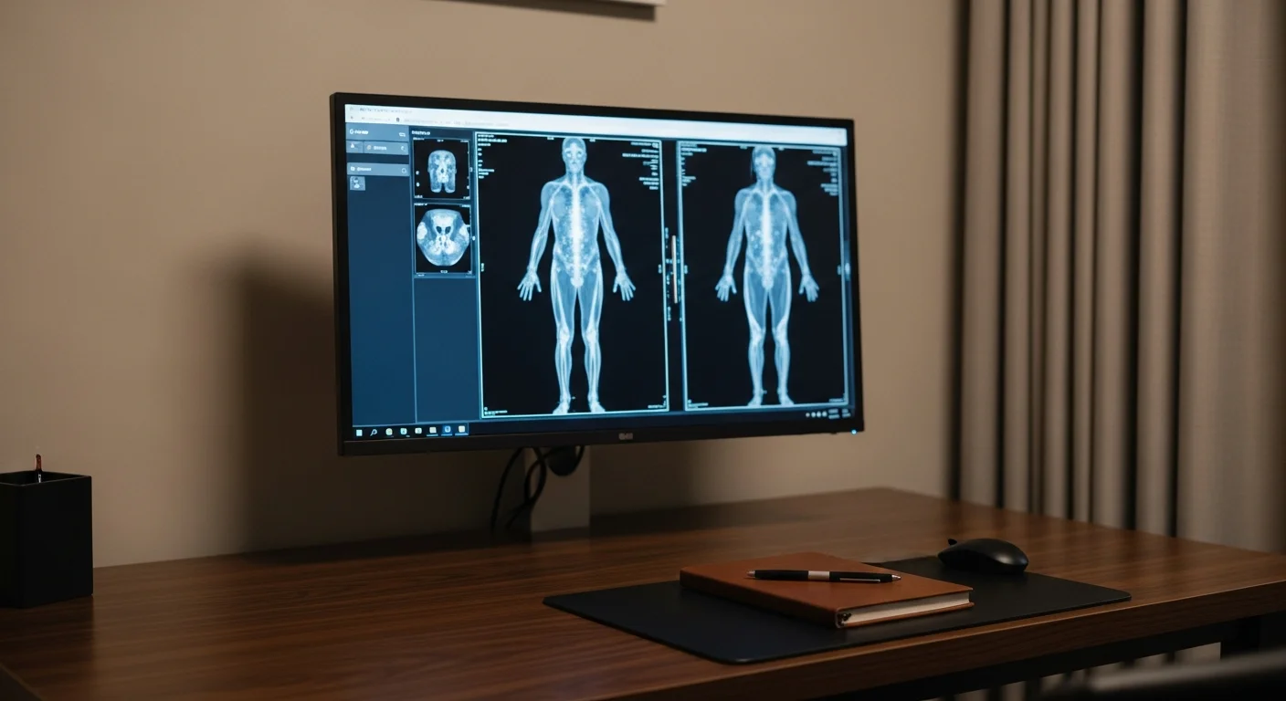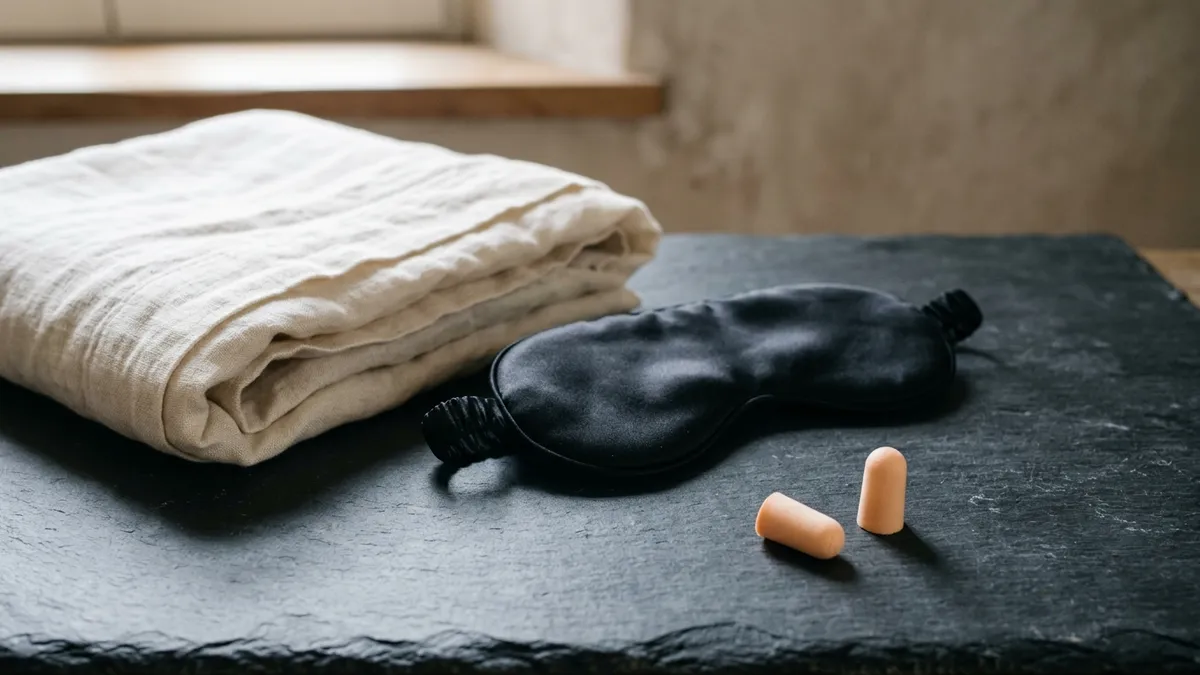
About 1.2% of MRI scans are stopped early because of claustrophobia, and the fix belongs before the appointment rather than on the table. Fishtown Medicine arranges a short-acting oral anxiolytic in hand with a driver, asks for a wide-bore scanner at standard field strength, and sorts out positioning, an eye mask, headphones, and a pause signal with the technologist.
TL;DR: If the MRI tube makes you panic, there is a plan for that, and it works far better when it is built before the day of the scan. About 1 in 100 MRI scans get stopped early because of claustrophobia, so imaging centers know this pattern well. Tell the doctor ordering the scan that the tube is a problem for you, and say it plainly if you have had to come out of one before. Ask for a short-acting anti-anxiety pill to have in hand ahead of time. It gets taken about an hour before you arrive and someone else has to drive you home, so it cannot be sorted out at the front desk on the day. Ask the imaging center for a wide-bore scanner, which is the roomier kind at full picture quality. Ask whether you can go in feet first or lie face down. Ask for an eye mask, headphones, and the squeeze ball that stops the scan. Ask how long the whole study takes and how long each noisy part lasts, so the sound stops being a surprise. Then book a time of day you know you can keep.
Coming out of a scanner partway through is a hard thing to describe to someone who has never had it happen. You went in willing, you meant to hold still, and somewhere in the middle your body decided it was done. What tends to stay with people afterward is less the fear itself and more the sense of having failed something other people get through without thinking about it.
That reading is unfair to you, and it also points at the wrong thing. Panic in a scanner is a predictable response to being held still in a loud, close space with your face a few inches from a wall, and the difference between a study that finishes and a study that stops is usually the set of arrangements made in the days before the appointment. What follows is what to ask for, in the order that matters, and what to do if you already have a stopped scan behind you.
How common is it to panic inside an MRI scanner?
Panic inside an MRI scanner is common enough that the published rates are consistent across countries and decades. A systematic review and meta-analysis in Radiography pooled 18 studies and found that claustrophobia terminated 1.18% of examinations.1 A cohort of 55,734 consecutive outpatients found claustrophobic reactions in 2.1% of people scanned on a conventional machine, falling to 0.7% on a newer machine that combined a short and wide bore with 97% acoustic noise reduction.2 A single-hospital review of 5,798 MRI reports found claustrophobia in 1.97% of patients, with 1.22% stopping the study because of it.3
Those percentages stay small until you multiply them out. The group behind the CLAUSTRO randomized trial estimated that roughly 2 million MRI examinations worldwide each year are either never completed or never attempted because of claustrophobia.4
The termination rate also undercounts what people go through. A review of 939 adults scanned over 7 weeks at a university hospital found that 14.3% needed oral sedation, intravenous sedation, or general anesthesia to tolerate the study.5 Add everyone who finishes while gripping the table the whole time, and this stops looking like a rare event and starts looking like a normal part of imaging that most centers already have a process for.
Predicting who will struggle is harder than it sounds. An 18-month study of 6,520 consecutive patients tested a 26-item Claustrophobia Questionnaire and found it useful for identifying who will be fine and limited when it came to picking out which individual would have trouble.6 The strongest single predictor is your own history. In a university hospital review of early MRI terminations, claustrophobia-related terminations occurred in 6.6% of people with a documented prior history of claustrophobia against 0.2% of everyone else.7
What does a stopped or blurred scan cost you?
A stopped scan costs more than the appointment slot. When an examination ends early, the sequences already captured are often unusable for the question that was asked, because MRI builds its answer out of several sequences that get read against one another. In that same university hospital series, 56% of early terminations were caused by claustrophobia, and terminations were more common among inpatients (1.2%) than outpatients (0.6%).7
Motion does the same damage more slowly. A review of a full week of clinical MRI at a single institution found significant motion artifact on sequences in 7.5% of outpatient examinations and 29.4% of inpatient and emergency department examinations, with repeat sequences run in 19.8% of all studies. The authors put the institutional cost at roughly $592 per hour of lost scanner time and about $115,000 per scanner per year.8 A separate review in Topics in Magnetic Resonance Imaging worked through the same arithmetic for claustrophobia, anxiety, and patient motion together.9
Your side of that ledger looks different from the hospital's. A repeat means another authorization, another day arranged around an appointment, another wait for a report, and a question about your health that stays open in the meantime. When the scan was ordered to settle something time-sensitive, the delay is the part that matters most, and it is the part nobody puts a price on.
What should I ask for before the appointment?
The request that changes the most is the one you make when the order is being written: tell whoever is ordering the scan that the scanner is a problem for you, and say plainly if you have had to come out of one before. Almost nothing below can be arranged at the front desk on the morning of the study, which is why the sentence has to come early.
Ask for these, in roughly this order:
- A prescription in hand before the day. A short-acting oral anxiolytic has to be taken before you arrive, so it needs to be written, filled, and explained in advance.
- A driver. Anyone who takes an oral anxiolytic before imaging needs another adult to drive them home afterward. Lining that person up is part of booking the appointment.
- The scanner details. Ask whether the center has a wide-bore scanner at 1.5T or 3T, and ask to be booked onto that machine.
- Feet-first entry, where the anatomy allows it. A knee, ankle, foot, and often a hip can be imaged with your head outside the bore.
- Prone positioning. Lying face down changes what you see, and it has been used to relieve claustrophobia in body MRI since 1984.10
- The length of the study and of each sequence. Ask how many minutes the whole thing runs and how long the individual sequences last.
- Comfort measures by name. An eye mask, prism glasses or a mirror, headphones with your own music, a blanket, and more airflow through the bore.
- A pause signal. Agree with the technologist what happens when you squeeze the ball, and whether you want to be talked to between sequences.
- A companion in the room. Many centers allow a screened family member or friend to stand beside the table with a hand on your ankle.
- A time of day you can keep. An evening or early-morning slot that you picked yourself survives a busy week better than one someone else booked for you.
How does premedication for an MRI work?
Premedication for an MRI usually means a single dose of a short-acting oral benzodiazepine taken 30 to 60 minutes before the scan, timed so the peak effect arrives while you are in the bore. The aim is anxiolysis, meaning enough softening of the fear response to let you hold still, rather than procedural sedation. You stay awake, you can talk to the technologist, and you still hear the noise.
Timing is where this most often goes wrong. A survey of adult outpatient MRI sedation practice found lorazepam was the first choice at 64% of centers and concluded that many outpatients are scanned before the peak effect of the drug they were given.11 A pill swallowed in the waiting room 10 minutes before the table is a pill doing very little by the time it counts.
The practical requirements are firm, and they are the reason this gets arranged in advance. Another adult drives you home; the ACR practice parameter for minimal and moderate sedation is explicit that a responsible adult accompanies an outpatient after discharge.12 You need to know the appointment time in order to know when to take the dose. And the prescriber needs your history first, including sleep apnea, other sedating medications, opioid or alcohol use, liver disease, pregnancy or breastfeeding, and your age, since older adults are more sensitive to these medications.
I am not printing doses here on purpose, because the right one depends on the person and belongs in a conversation with a clinician who knows your history. What I want you to know is that asking for it is ordinary. It is among the most common requests in outpatient imaging, and the person on the other end of that request hears it every week.
Oral premedication has limits worth understanding before you rely on it. It softens the response; it does not remove the noise, the confinement, or the requirement to hold still. When it proves insufficient, the next steps are intravenous sedation with monitoring or an anesthesia-assisted scan, and both have to be booked as such rather than improvised on the day. One Mayo Clinic series found that moving claustrophobic patients onto a 1.5T wide short-bore scanner raised the examination success rate and substantially reduced the need for anesthesia-assisted MRI even when the claustrophobia was severe,13 which is why the scanner question deserves to come before the anesthesia question.
What helps once you are on the table?
Several non-drug measures have evidence behind them, and they work together rather than competing. A systematic review of 21 studies of adults undergoing MRI grouped the approaches that helped into scanner design features, cognitive and behavioural strategies, prone positioning, information provision, fragrance, and team training, and found some positive effect across all of them.14
Prone positioning is the oldest of these. Lying face down lets you look toward the table and the floor instead of up at a surface a few inches from your face, and it was described in Radiology as a way to relieve claustrophobia in body MRI back in 1984.10 It is available for many body and spine studies and unavailable for others, so ask rather than assume.
Feet-first entry does something simpler: it keeps your head outside the bore. A knee, ankle, foot, and often a hip can be scanned that way. A brain, cervical spine, shoulder, or chest study cannot, because the region being imaged has to be at the center of the magnet, and that geometry is fixed by physics rather than by preference.
Being able to see out of the tube helps a great many people, which is why mirrors and prism glasses are standard equipment at most centers. A randomized trial published in Radiography enrolled 226 people with known claustrophobia, about half of whom had been unable to complete a previous MRI, and compared person-centred nursing care alone against the same care plus immersive video glasses and headphones. Across the trial, 83.6% completed the scan.15
Preparation is its own intervention. In a 2023 study of claustrophobic patients describing their own experiences, the themes that emerged were examination preparation, information provision, coping mechanisms, and scanner design.16 Knowing that the next sequence runs 4 minutes and sounds like a jackhammer turns a frightening surprise into a countdown you can ride out.
Rehearsing calm before you arrive has trial support too. An observational 2-group study using an audio-guided self-hypnosis track in the waiting room found claustrophobic events in 16% of the self-hypnosis group (9 of 55) against 43% of the control group (38 of 89), with sedation needed in 2% against 16%.17 Slow breathing practised in advance works on the same principle and costs nothing.
The squeeze ball earns its place. Every scanner has a ball or call button wired to an alarm in the control room, and the scan stops when you use it. Decide with the technologist beforehand what happens next: whether they talk to you and continue, pause and restart the sequence, or bring you out. Knowing you hold that switch changes the feeling of the bore considerably.
Fishtown Medicine
A 90-minute conversation with Dr. Ash. A written plan you can follow.
Headphones are standard for hearing protection, and most centers will play your own music through them. The trial evidence for music by itself during MRI is thinner than the evidence for positioning and preparation, so treat it as a low-cost addition rather than the plan. An eye mask belongs in the same category: widely offered, without trial data I can point you to, and helpful for many people because it removes the visual information about how close the bore is. Putting it on before you are moved into the magnet is the step people forget.
Is an open MRI the answer?
Open MRI is the option people ask for by name, and the answer is that it helps less than most people expect and costs more image quality than most people realize.
The CLAUSTRO randomized controlled trial assigned 174 patients to an open scanner or a short-bore closed scanner. Claustrophobic events occurred in 26% of the open group and 39% of the short-bore group, a difference that did not reach statistical significance (P = 0.08).18 Open design reduced events somewhat and did not remove them.
The companion randomized comparison of image quality is where the trade-off becomes visible. In that trial, 93 patients had spine MRI on either a 1.0T open scanner or a 1.5T short-bore scanner. Overall qualitative image quality was 39% higher on the short-bore machine, signal-to-noise ratio was 53% higher, and quantitative contour sharpness was 34% to 37% higher. Scans also took longer on the open unit, averaging 32 minutes against 20.19 No sequence came out non-diagnostic, so open MRI produces usable images; it produces less detailed ones and takes longer doing it.
Wide-bore at standard field strength is usually the better compromise, because it keeps 1.5T or 3T image quality while giving you a wider opening and, on newer units, much less noise. The 55,734-patient cohort found claustrophobic reactions in 0.7% of scans on a short and wide-bore machine with acoustic noise reduction against 2.1% on the conventional scanner, an adjusted odds ratio of 3.1.2 The Mayo Clinic series above found the same pattern in people whose claustrophobia was already severe.13
So the order to ask in is wide-bore at standard field strength first, then open MRI, and open MRI mainly when the clinical question tolerates lower resolution or when nothing else will get you scanned at all. For a question that hinges on fine detail, such as a small brain lesion or cartilage, the resolution you give up on an open unit can cost you the answer you came for.
Can the scan be shortened, or done a different way?
Some scans can be shortened, though not on request and not everywhere. Abbreviated protocols are validated in specific settings rather than offered as a general convenience. Abbreviated breast MRI is the clearest example: the EA1141 trial randomized 1,516 women with dense breasts, used an abbreviated protocol acquired in under 10 minutes, and detected considerably more cancers than digital breast tomosynthesis.20 Abbreviated liver MRI for hepatocellular carcinoma surveillance is built the same way, and biparametric prostate MRI drops the contrast-enhanced portion. Outside settings like these, a radiologist can sometimes trim a protocol down to the specific question, or split a long study across 2 visits. Either one is a conversation between the ordering clinician and the radiologist rather than something a scheduler can approve.
Substituting a different test deserves the most caution of anything on this page. For some questions, CT or ultrasound gives an adequate answer: acute bleeding in the head, a skull fracture, kidney stones, gallbladder disease, many superficial soft-tissue questions, and most abdominal emergencies. For other questions the substitution costs the diagnosis outright. CT cannot show multiple sclerosis plaques, early spinal cord compression, or marrow swelling in a stress fracture that has yet to appear on X-ray, and ultrasound cannot see through bone or gas. Trading the test that answers your question for the test you can tolerate buys you a calmer hour today and leaves the question open for months. When the substitute does answer the question, taking it is a good decision; when it does not, the better plan is to make the MRI possible. Why we choose an MRI works through which questions each study can settle.
What if you already had to stop a scan partway through?
Start with the sentence that does the most work: when you book the repeat, say that you had to come out of a previous scan. That fact moves you onto a different pathway at most imaging centers, and it earns its weight, since a documented history of claustrophobia was associated with early termination in 6.6% of examinations against 0.2% without one.7 You are handing them something they can act on rather than a complaint.
Then ask what was already captured. An examination that ended early is not always a total loss, and sometimes enough sequences were acquired to answer the question, or to narrow the repeat down to 2 or 3 targeted sequences instead of the whole protocol. Ask the ordering clinician or the radiologist which sequences are missing, what the repeat has to cover, and whether contrast will be needed again.
Then rebuild the appointment around what went wrong the first time. If the fear peaked as your head entered the bore, feet-first entry, prone positioning, or a wide-bore machine addresses that directly. If it built slowly through the study, premedication and a sequence-by-sequence countdown matter more. If the noise started it, ask about a scanner with acoustic noise reduction, better ear protection, and a technologist who will talk to you between sequences.
And give yourself an appointment you can keep. A slot you chose yourself, on a day you are not sprinting there from work, with the driver already arranged, is a different event from one that was assigned to you.
How Fishtown Medicine handles MRI anxiety in Philadelphia
When Fishtown Medicine orders an MRI for someone who has had trouble in a scanner, the anxiolytic prescription goes out alongside the imaging order rather than after a failed attempt, with the driver requirement explained and the timing written down. The imaging order goes directly to the patient as well as to the center, so the appointment can be self-scheduled into an evening or early-morning slot that will survive a busy week. The appointment you can keep is the one that gets done.
We ask which scanner before booking, and we tell the center in advance that claustrophobia is part of the picture so it is in the notes before you arrive rather than a surprise at the table. For patients paying cash, independent imaging centers around Philadelphia often quote a fraction of the hospital-affiliated price for the same study, and the wide-bore question is worth asking of each one.
The report then comes with a conversation. An MRI report lists everything the radiologist sees whether or not it bears on your symptom, and read alone at 11 PM it is frightening. What you need from it is which finding explains what brought you in, which findings are ordinary for your age, and what happens next.
Guidance from the Clinic

Actionable Steps for Philly Patients Facing an MRI
Work through these in order, starting the day the scan is ordered.
- Say it when the order is written. Tell the ordering clinician that the scanner is a problem for you, and mention any scan you have had to stop before.
- Get the prescription in hand and know the timing. Ask what to take, how long before arrival to take it, and what to avoid alongside it.
- Arrange the driver before you book the slot. Another adult has to take you home, so pick the appointment around their schedule too.
- Ask the imaging center 3 things. Whether the scanner is wide-bore, how many minutes the full study runs, and whether feet-first entry works for your body part.
- Ask for the comfort measures by name. Eye mask, prism glasses or mirror, your own music through the headphones, a blanket, more airflow, and a companion in the room.
- Agree a pause signal with the technologist. Decide together what happens when you squeeze the ball, and ask them to call out the length of each sequence.
- Book a slot you can keep. An evening or early-morning appointment you chose yourself beats a mid-afternoon one you will be tempted to cancel.
Key Takeaways
- Claustrophobia terminates about 1.2% of MRI examinations, and roughly 2 million scans worldwide each year are never completed or never attempted because of it, so this is a recognized problem with a worked-out response.
- The single most useful thing you can do is tell the ordering clinician and the imaging center in advance, since a documented prior history predicts early termination at 6.6% against 0.2%.
- A short-acting oral benzodiazepine taken 30 to 60 minutes before the scan is a standard approach, and it must be arranged ahead of time because the dose is taken before arrival and another adult has to drive you home.
- Prone positioning, feet-first entry where the anatomy allows, mirrors or prism glasses, an eye mask, headphones, a companion in the room, and a rehearsed pause signal all help, and preparation is itself an intervention.
- Wide-bore at 1.5T or 3T beats open MRI as a first ask: a randomized trial found no significant claustrophobia advantage for open scanners, while a companion trial found image quality 39% lower and scan times longer on the open unit.
- Abbreviated protocols exist where they have been validated, such as breast MRI and liver surveillance, and swapping in a CT or ultrasound that cannot answer the question trades a calmer hour today for months of uncertainty.
Related at Fishtown Medicine
- Why We Choose an MRI, and When We Do Not - what MRI settles, when it is the wrong first study, and how implants are handled
- Why We Choose an MRA, Territory by Territory - imaging arteries without radiation, artery group by artery group
- Understanding Brain and Head Imaging - how CT and MRI divide neurologic questions
- When You Need an Ultrasound - the radiation-free option for superficial structures and flow
- When to Order Imaging - the decision framework behind every scan
- Boutique Scans and Whole-Body MRI - why screening someone without symptoms is a different problem
Scientific References
- Munn Z, Moola S, Lisy K, Riitano D, Murphy F. "Claustrophobia in magnetic resonance imaging: a systematic review and meta-analysis." Radiography. 2015;21(2):e59-e63. Radiography
- Dewey, Schink, and Dewey. "Claustrophobia during magnetic resonance imaging: cohort study in over 55,000 patients." Journal of Magnetic Resonance Imaging. 2007;26:1322-1327. Wiley
- Eshed et al. "Claustrophobia and premature termination of magnetic resonance imaging examinations." Journal of Magnetic Resonance Imaging. 2007. PubMed
- Enders J, Zimmermann E, Rief M, et al. "Reduction of claustrophobia during magnetic resonance imaging: methods and design of the CLAUSTRO randomized controlled trial." BMC Medical Imaging. 2011;11:4. BMC Medical Imaging
- Murphy and Brunberg. "Adult claustrophobia, anxiety and sedation in MRI." Magnetic Resonance Imaging. 1997. PubMed
- Napp, Enders, Roehle, et al. "Analysis and prediction of claustrophobia during MR imaging with the Claustrophobia Questionnaire: an observational prospective 18-month single-center study of 6500 patients." Radiology. 2017;283(1):148-157. PubMed
- "Early MRI termination with major impact on the radiological interpretation: the experience of a large university hospital." European Journal of Radiology. 2023. PubMed
- Andre et al. "Toward quantifying the prevalence, severity, and cost associated with patient motion during clinical MR examinations." Journal of the American College of Radiology. 2015. PubMed
- Nguyen XV, Tahir S, Bresnahan BW, et al. "Prevalence and financial impact of claustrophobia, anxiety, patient motion, and other patient events in magnetic resonance imaging." Topics in Magnetic Resonance Imaging. 2020;29(3):125-130. Topics in Magnetic Resonance Imaging
- Hricak H, Amparo EG. "Body MRI: alleviation of claustrophobia by prone positioning." Radiology. 1984;152(3):819. PubMed
- Middelkamp JE, Forster BB, Keogh C, Lennox P, Mayson K. "Evaluation of adult outpatient magnetic resonance imaging sedation practices: are patients being sedated optimally?" Canadian Association of Radiologists Journal. 2009. Canadian Association of Radiologists Journal
- American College of Radiology and Society of Interventional Radiology. "ACR-SIR Practice Parameter for Minimal and/or Moderate Sedation/Analgesia." ACR
- Hunt CH, Wood CP, Lane JI, Bolster BD, Bernstein MA, Witte RJ. "Wide, short bore magnetic resonance at 1.5 T: reducing the failure rate in claustrophobic patients." Clinical Neuroradiology. 2011;21(3):141-144. PubMed
- Munn Z, Jordan Z. "Interventions to reduce anxiety, distress and the need for sedation in adult patients undergoing magnetic resonance imaging: a systematic review." International Journal of Evidence-Based Healthcare. 2013. PubMed
- "Claustrophobia in magnetic resonance imaging: a randomised controlled trial of immersive entertainment glasses." Radiography. 2026;32(2):103318. Radiography
- "Supporting claustrophobic patients during magnetic resonance imaging examination: the patient perspective." Radiography. 2023. PubMed
- "Audio-guided self-hypnosis for reduction of claustrophobia during MR imaging: results of an observational 2-group study." European Radiology. 2021. PubMed
- "Reduction of claustrophobia with short-bore versus open magnetic resonance imaging: a randomized controlled trial." PLoS One. 2011;6(8):e23494. PubMed
- "High-field open versus short-bore magnetic resonance imaging of the spine: a randomized controlled comparison of image quality." PLoS One. 2013;8(12):e83427. PubMed
- Comstock et al. "Comparison of abbreviated breast MRI vs digital breast tomosynthesis for breast cancer detection among women with dense breasts undergoing screening." JAMA. 2020. PubMed

Frequently Asked Questions
Common Questions
Deep-Dive Questions
Ready when you are
The chat is our AI assistant, answering from our published guides. To talk it through with Dr. Ash himself, start with the intake.